Cartilage Restoration Surgeon in Annapolis, Maryland
Cartilage is the one tissue in the knee that does not heal. A pothole in the smooth surface of the joint stays a pothole, and in an active person it tends to get larger. Cartilage restoration is the group of operations that fill that pothole, with the goal of relieving pain and keeping a young, active knee out of the arthritis clinic for as long as possible.
Dr. Benjamin M. Petre is a board-certified orthopedic surgeon, fellowship-trained in sports medicine at The Steadman Clinic in Vail, Colorado, where modern microfracture was developed. He performs cartilage restoration surgery at Luminis Health Orthopedics in Annapolis, including microfracture, osteochondral autograft and allograft transplantation, and cell-based repair, as part of about 150 knee operations a year. Patients come from Annapolis, Anne Arundel County, and across Maryland.
These are operations for focal cartilage damage: a defined defect in an otherwise healthy joint. They are not treatments for arthritis, where the cartilage is worn thin across the whole joint. Dr. Petre does not perform joint replacement, but he has excellent partners in Luminis Health's joint replacement program and is glad to refer you to them. Part of a cartilage evaluation is an honest answer about which of the two you have.
Anatomy: What Articular Cartilage Does
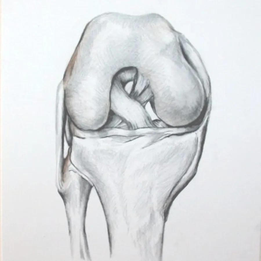
Articular cartilage is the smooth, white layer, a few millimeters thick, that caps the ends of the bones inside a joint. In the knee it covers the end of the femur, the top of the tibia, and the back of the kneecap. It is slicker than ice on ice and spreads load across the bone beneath it.
It is also unusual tissue. It has no blood supply and no nerves, so it cannot mount a healing response and it does not hurt directly. Pain comes later, from the overloaded bone under a defect, from swelling, and from loose fragments. That is why a cartilage injury can be silent for a while and why it does not repair itself the way a muscle or a bone does.
The knee has two types of cartilage. Articular cartilage lines the ends of the bones, and it is a different tissue from the meniscus. The menisci are two rubbery, C-shaped shock absorbers, made of a tougher type called fibrocartilage, that sit between the bones. The two are often injured together, and a knee that has lost meniscus puts more load on the articular cartilage it has left.
What Is a Cartilage Defect?
A cartilage defect is an area where the surface is damaged or missing. Surgeons grade it by depth, from softening and surface fraying to a full-thickness hole down to bone, and measure it by size and location. Those three things, along with your age and the state of the rest of the knee, decide the treatment.
Two related conditions belong in the same conversation:
- Osteochondral defects involve the cartilage and the bone beneath it, often after a dislocation of the kneecap or a hard impact.
- Osteochondritis dissecans (OCD) is a condition, usually starting in adolescence, in which a segment of bone under the cartilage loses its blood supply and the piece can loosen or break free.
What a defect is not is arthritis. In arthritis the cartilage is thin everywhere, the joint space narrows on X-ray, and there is no healthy shoulder of cartilage around the damage to contain a repair. Restoration procedures do poorly in that setting, and it would be a disservice to offer them.
Causes
- A single injury. A twist, a direct blow, or a dislocation of the kneecap can shear cartilage off the bone. Cartilage damage is common alongside ACL tears and patellar dislocations.
- Repetitive overload. A knee that is unstable, out of alignment, or missing meniscus concentrates force on one area until the cartilage fails.
- Osteochondritis dissecans.
- Previous surgery. Removal of a significant amount of meniscus years earlier is a common backstory.
Symptoms
- Pain with weight-bearing, often localized to one side of the knee or behind the kneecap, and worse with stairs, squatting, or impact.
- Swelling that comes on after activity and settles with rest.
- Catching, locking, or a sense that something is moving inside the knee, which suggests a loose fragment.
- A knee that feels fine walking and fails when you try to run or cut.
A locked knee that will not straighten, or a large swelling after an injury, should be evaluated promptly. A displaced fragment is far easier to fix early than late.
Diagnosis
Evaluation starts with the story and an examination of alignment, kneecap tracking, ligament stability, and where the knee is tender. X-rays, including standing views and often a full-length alignment film from hip to ankle, show joint space and whether the leg is bow-legged or knock-kneed. MRI maps the defect: its size, depth, location, and the condition of the bone beneath it, the menisci, and the ligaments.
MRI tends to underestimate cartilage damage, so the definitive measurement is often made with an arthroscope. For some of the procedures below, that first arthroscopy is also when a small cartilage sample is taken to grow cells for a later repair.
The findings that matter are not only in the defect. A cartilage repair placed into a knee that is unstable, malaligned, or missing its meniscus is being asked to survive the same forces that caused the damage. Finding and correcting those problems is as much a part of cartilage restoration as the repair itself.
Non-Surgical Treatment
Many cartilage defects are found by accident on an MRI and never cause trouble. A defect that is small, shallow, or not causing symptoms is watched, not operated on. For symptomatic defects, a fair trial of non-surgical care comes first in most cases: activity modification, physical therapy for quadriceps and hip strength, weight management where relevant, unloading braces, and injections to settle swelling and pain.
None of these regrow cartilage. They reduce symptoms and load, and for many people that is enough. Dr. Petre and his PA and NP team provide this care in the same office.
Who Is a Candidate for Cartilage Restoration?
The best candidates share a profile:
- A focal, full-thickness or near full-thickness defect that matches the symptoms.
- Healthy cartilage around the defect and on the opposite surface.
- An age and activity level where preserving the joint matters. Most patients are between their teens and their forties.
- A knee that is stable and well aligned, or one where instability, alignment, and meniscus loss can be corrected at the same time.
- Willingness to commit to a long, restricted rehabilitation. These repairs are won or lost in the first months.
Poor candidates are just as important to identify: diffuse arthritis, defects on both sides of the joint facing each other, inflammatory arthritis, significant obesity, and nicotine use all lower the odds of success. When restoration is not the right tool, Dr. Petre says so and helps arrange the right referral.
Cartilage Restoration Procedures
There is no single best cartilage operation. The choice depends on the size and depth of the defect, whether the bone beneath it is involved, your age, and what you want to return to.
Chondroplasty
For partial-thickness damage with unstable flaps, the loose edges are smoothed arthroscopically to stop them from catching. It is the smallest operation and has the quickest recovery, and it does not fill the defect.
Microfracture
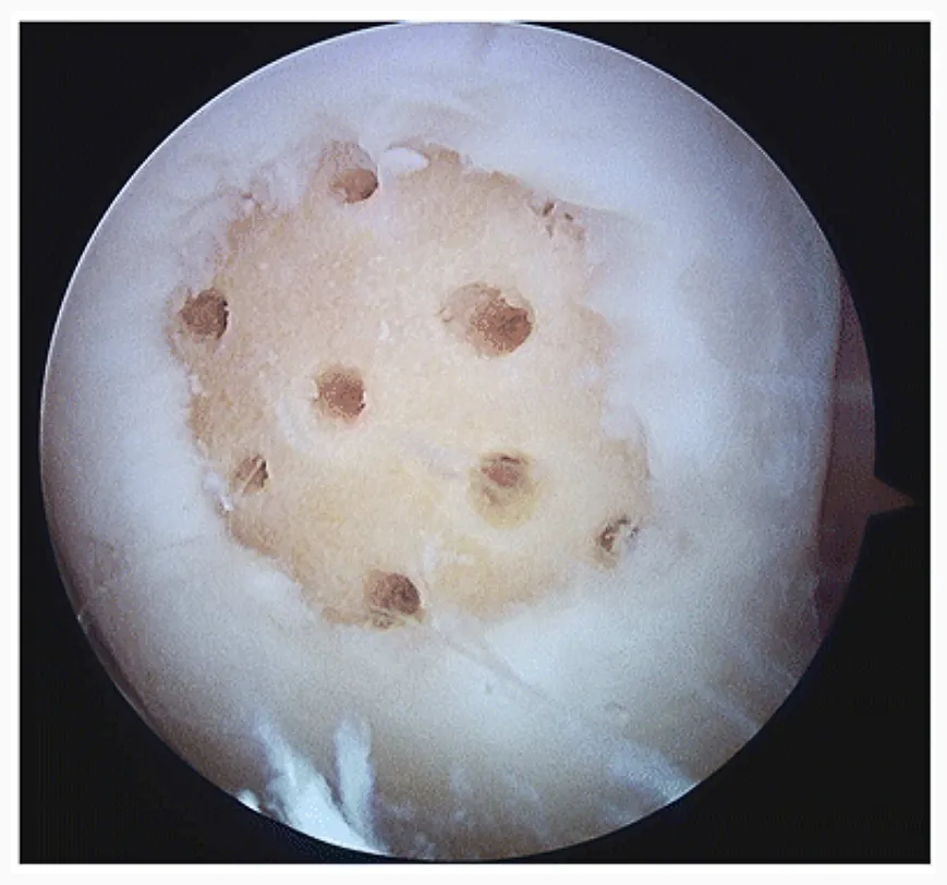
Small holes are made in the bone at the base of the defect, releasing marrow cells that form a clot and mature into a repair tissue called fibrocartilage. It is arthroscopic, done in a single operation, and works best for small defects in younger patients. The repair tissue is not as durable as native cartilage, which is why it is reserved for smaller lesions.
Osteochondral autograft transfer (OATS)
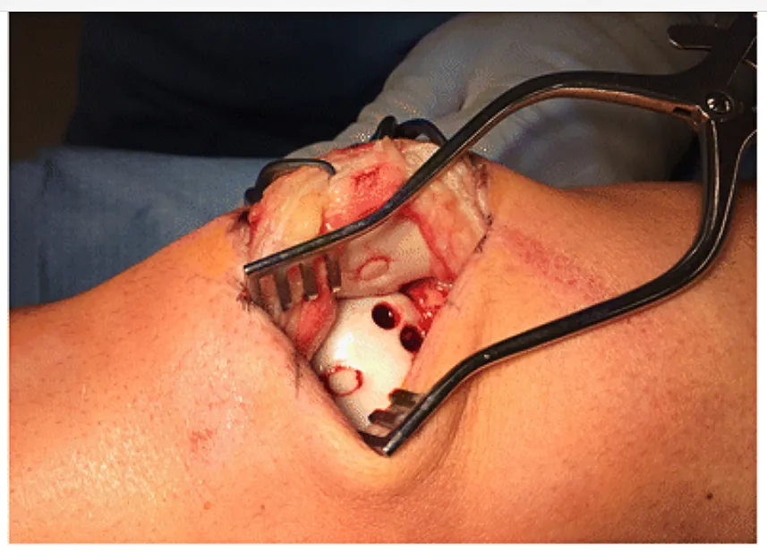
One or more cylindrical plugs of healthy cartilage and bone are taken from a low-load area at the edge of your own knee and press-fit into the defect. The repair is real hyaline cartilage on living bone, and it heals bone-to-bone. It suits small to medium defects, particularly in high-demand athletes.
Osteochondral allograft transplantation (OCA)
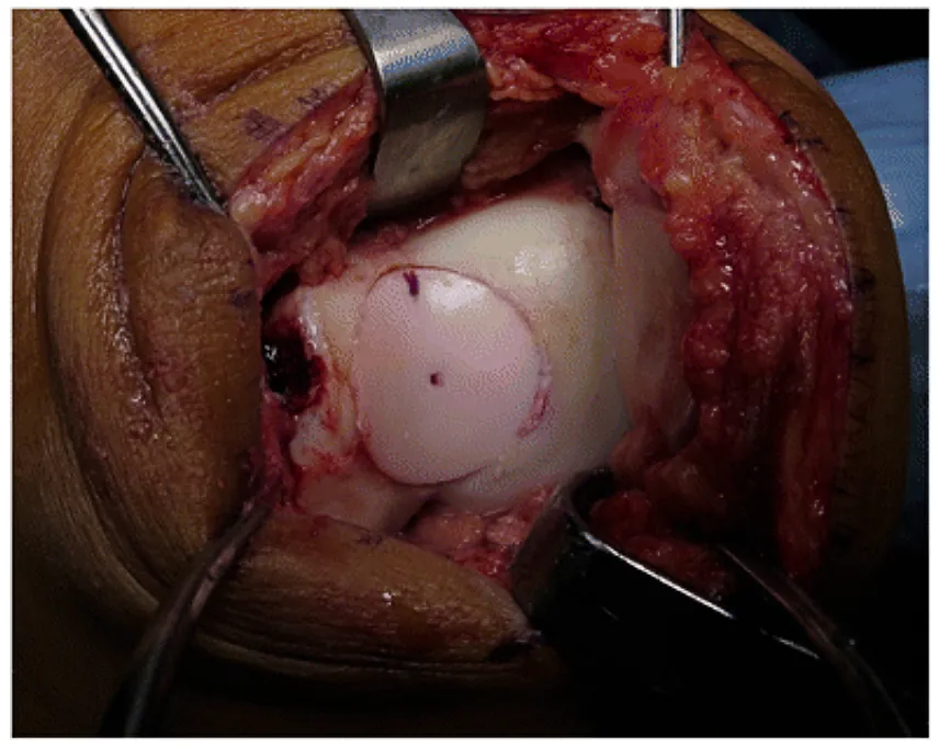
For larger defects, or defects that involve the bone, a size-matched plug of fresh cartilage and bone from a donor is transplanted into the defect. It restores a large area with mature cartilage in one operation and is a workhorse for failed prior repairs and for osteochondritis dissecans. Surgery is scheduled around the availability of a matched graft.
MACI (matrix-induced autologous chondrocyte implantation)
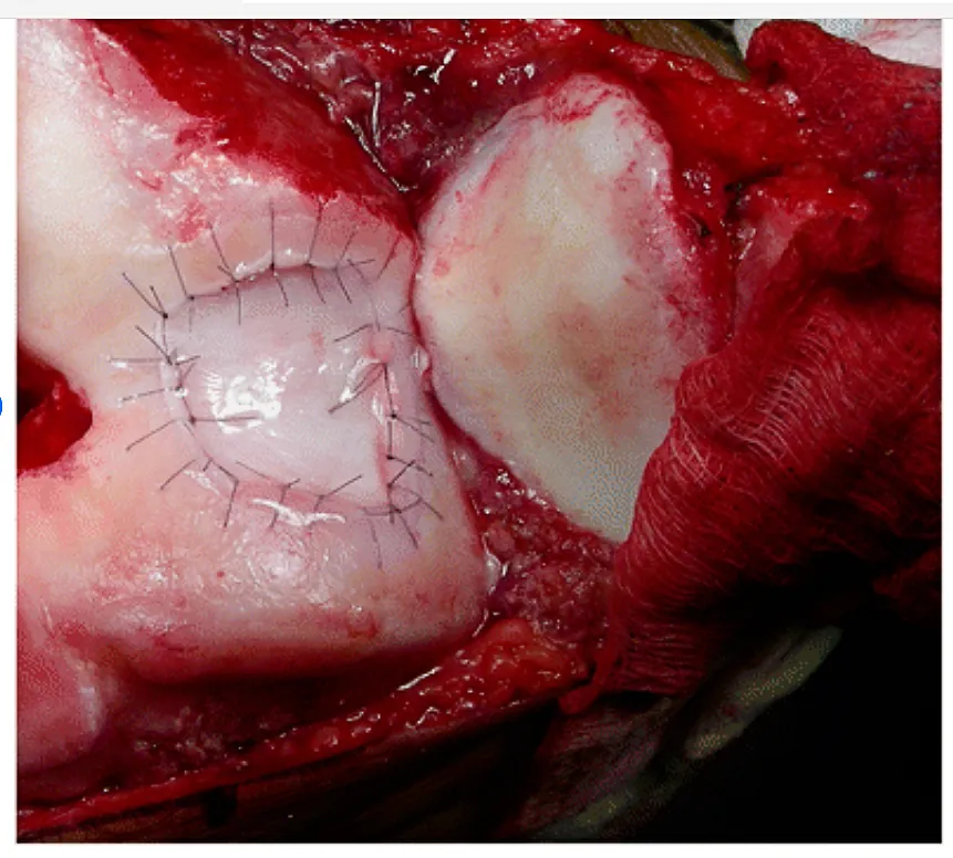
A two-stage procedure. At a first arthroscopy, a small sample of your cartilage is harvested and sent to a laboratory, where the cells are grown on a collagen membrane over several weeks. At a second operation the membrane is trimmed to shape and glued into the defect. MACI suits medium to large defects where the bone is intact, including defects behind the kneecap.
Procedures that protect the repair
Cartilage repairs fail when the forces that caused the damage are left in place. Depending on your knee, the same operation may include an osteotomy to shift load away from the repaired side, a meniscus transplant, ligament reconstruction, or realignment of the kneecap.
Cartilage damage in the hip is treated differently, during hip arthroscopy.
Preparing for Surgery
- Stop nicotine in every form. It impairs graft and bone healing.
- Plan for crutches. Most cartilage repairs require four to eight weeks of little or no weight on the leg, which affects driving, work, stairs, and childcare.
- Set up physical therapy before surgery day.
- Hold anti-inflammatories and blood thinners as directed in your pre-op instructions.
- Review our preparing for major knee surgery checklist for what to have at home.
The Procedure, Step by Step
- Anesthesia. General anesthesia, usually with a regional nerve block for the first day's pain.
- Arthroscopy. The whole knee is inspected. The defect is measured, loose fragments are removed, and the menisci and ligaments are assessed.
- Defect preparation. Damaged cartilage is removed back to a stable, healthy rim with vertical walls. Good preparation is common to every technique.
- Restoration. Microfracture and chondroplasty are completed through the arthroscope. Plug transfers, allograft transplants, and MACI are placed through a short incision beside the kneecap so the graft can be seated flush with the surrounding surface.
- Associated procedures. Any osteotomy, meniscus, or ligament work is done in the same operation where possible.
- Closure. The knee is dressed and placed in a brace. Most patients go home the same day.
Recovery and Return to Sport
Recovery from cartilage restoration is long, and it is strict early. The graft or repair tissue is soft at first and matures over many months. The timelines below follow our published protocols and vary with the procedure, the location of the defect, and anything else done at the same time. Your post-op instructions are the final word.
Weeks 0 to 6
Crutches, with little or no weight on the leg: about six weeks after microfracture, about four weeks before a gradual return after cell-based repair, and about eight weeks after an allograft transplant. A brace is locked straight for walking until the quadriceps is firing well. Therapy starts in the first week with quadriceps activation, kneecap mobility, and range of motion within the limits set for your defect.
Weeks 6 to 12
Weight-bearing is restored, crutches are put away, and gait is retrained. Stationary cycling adds resistance, and closed-chain strengthening begins within protected ranges of knee bend that depend on where the defect is.
Months 3 to 6
Progressive strengthening, single-leg work, and low-impact conditioning such as the elliptical and swimming. A running progression typically begins somewhere in this window, often on an unweighting treadmill first, and only when strength and swelling allow.
Beyond six months
Return to cutting and impact sport is generally six to nine months after microfracture and OATS and nine to twelve months or more after allograft transplantation and MACI, decided by functional testing, not the calendar. Some high-impact activity may not be advisable after the largest reconstructions, and that conversation happens before surgery, not after.
The full schedules are in our microfracture, cartilage repair, and osteochondral allograft protocols, written for you and your physical therapist.
Risks and Complications
Possible complications include infection, bleeding, blood clot, stiffness, and anesthesia risks. Specific to cartilage surgery: the repair may not fill completely or may not integrate with the surrounding cartilage; repair tissue after microfracture can wear over time; a transplanted plug can fail to heal to the bone or can collapse; MACI tissue can overgrow and catch; and plug harvest for OATS can leave soreness at the donor site. Stiffness is the most common problem early and is the reason motion starts on day one. Some patients need a further operation.
Cartilage restoration improves pain and function for most well-selected patients, but it does not make a knee new, and no surgeon can guarantee an outcome. What you can expect is a candid assessment of whether your knee is a good candidate, and which procedure gives it the best odds.
Frequently Asked Questions
Can knee cartilage heal on its own?
No. Articular cartilage has no blood supply, so a full-thickness defect does not fill in by itself. Symptoms can still settle with therapy, bracing, and activity changes, and many defects never need surgery. When symptoms persist in an active person, restoration procedures are designed to fill the defect.
Is cartilage restoration the same as treating arthritis?
No. Restoration procedures repair a defined area of damage in an otherwise healthy joint. Arthritis is thinning of cartilage across the whole joint, and these operations do poorly in that setting. Dr. Petre does not perform joint replacement, but he can refer you to excellent partners in Luminis Health's joint replacement program, and the evaluation will tell you which problem you have.
What is the difference between microfracture, OATS, and MACI?
Microfracture stimulates the bone to form repair tissue and suits small defects. OATS moves plugs of your own cartilage and bone into the defect and suits small to medium defects. MACI grows your own cartilage cells on a membrane that is implanted at a second operation, and suits medium to large defects. Osteochondral allograft uses donor cartilage and bone for the largest defects and those involving bone.
How long will I be on crutches after cartilage surgery?
Usually four to eight weeks with little or no weight on the leg, depending on the procedure: about six weeks after microfracture and about eight weeks after an allograft transplant. Protecting the repair in this period is the single most important thing you can do for the result.
When can I run or play sports after cartilage restoration?
A running progression typically begins between three and six months when strength and swelling allow. Return to cutting and impact sport is generally six to nine months after microfracture or OATS and nine to twelve months or more after allograft or MACI, decided by functional testing.
Am I too old for cartilage restoration?
There is no single cutoff. Results are best in patients from their teens through their forties with a focal defect and healthy surrounding cartilage. What matters more than age is whether the damage is a contained defect or generalized arthritis.
Will I need other procedures at the same time?
Often. If the leg is out of alignment, the knee is unstable, or meniscus is missing, the same forces that damaged the cartilage will damage the repair. Correcting them with an osteotomy, ligament reconstruction, or meniscus transplant at the same operation gives the repair its best chance.
Does insurance cover cartilage restoration?
Most plans cover these procedures when criteria are met, and several of them, including MACI and allograft transplantation, require pre-authorization. Our surgery coordinator handles that process before a date is set. Call 410-280-4716 with authorization questions.
Get Your Knee Cartilage Evaluated in Annapolis
Luminis Health Orthopedics, 2000 Medical Parkway, Suite 101, Annapolis, MD 21401. Monday–Friday, 8:00 AM–5:00 PM.
Related: ACL Reconstruction·MPFL Reconstruction & Patellar Instability·Hip Arthroscopy·Knee Conditions·Preparing for Knee Surgery·Rehabilitation Protocols·About Dr. Petre
Dr. Petre's work is focused exclusively on sports medicine and sports injury surgery. He does not perform joint replacement, but he has excellent partners in Luminis Health's joint replacement program and is glad to refer you to them.
This page is provided for general education and is not a substitute for individual medical advice. The right treatment depends on your examination, imaging, and goals. Please consult Dr. Petre or another qualified physician about your specific condition.
